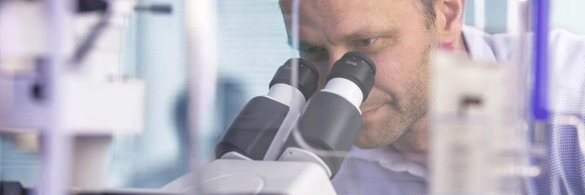

나에게 필요한 광학 현미경 찾기
우리 학교, 연구실, 기관을 위한 ZEISS 광학 현미경 솔루션


정립·도립 현미경 어플리케이션 미리보기
-
HEK 293 Cells
Long-term time lapse recording of 3 × 3 tiles with 240 s interval. Acquired with Axiocam 506 mono, stabilized by Definite Focus 3 at 10 s interval.
HeLa Cell Culture
HeLa cell culture with cytosolic eGFP. Proliferation imaged over 16 hours.


SK8 K18 Mouse Cells
Vimentin stained with Alexa 488 (green), nuclei stained with DAPI (blue).


Cortical Neurons
Cortical neurons stained for DNA, microtubules and microtubule-associated proteins. Courtesy: Leibniz-Institute on Aging – Fritz-Lipmann-Institut e.V. (FLI), Germany.
LLC PK1 Cells
Time lapse recording of undisturbed cell division.


Spherulitic graphite in nodular cast iron, reflected light, brightfield


Cast aluminum-silicon, reflected light, darkfield


Barker-etched aluminum, reflected light, C-DIC


Zinc, reflected light, polarization contrast with lambda plate


Niccolite, reflected light, polarization contrast with lambda plate
-


Histological specimen (human)


Chromosome specimen (human)


Renal tissue (human)


Mink uterus endometrium epithelial cells


Indian muntjac, fibroblasts


Mouse kidney in fluorescence


Novicula lyra


Crystal


Trout cartilage
-


Blood vessels, transmitted light brightfield


Red bone marrow human in transmitted light brightfield


Blood smear, Giemsa staining, transmitted light brightfield


Rat retina, section, nuclear fast red, transmitted light brightfield


Rat tongue, acid green, transmitted light brightfield

Varroa mite, transmitted light brightfield


Representative image of human blood showing leukemia, Giemsa staining, transmitted light brightfield


Cast iron, reflected light, bright field, EC EPIPLAN 20×/0.4


Cast iron, reflected light, dark field, EC EPIPLAN 20×/0.4


Electronic device SMD resistor: Document the quality of printing and contact surfaces. EC EPIPLAN 5×/0.13


Injection-molded identification letter at transparent plastic part, EC EPIPLAN 5×/0.13


Milling marks for engraving purposes, EC EPIPLAN 5×/0.13


Biotite inside granite, transmitted light, bright field, EC Plan-NEOFLUAR 10×/0.3 Pol
-


High-throughput TMA analysis: 13 tissue stamps were screened using a set of seven markers for tumor analysis to gain insights into tumor-immune interactions and find potential therapeutic targets


Fixed U2OS cells with stained lysosomes (yellow), DNA (blue), and actin (magenta)


Fixed human tonsil sample stained for DNA (blue), panCK (green), CD3 (Orange), PD-L1 (red), and CD68 (purple)


Sample courtesy of Annekatrin Richter, Leibniz Institute in Aging, Fritz Lipmann Institute e.V. (FLI), Jena, Germany. Sample courtesy of Annekatrin Richter, Leibniz Institute in Aging, Fritz Lipmann Institute e.V. (FLI), Jena, Germany. Hematoxylin and eosin-stained section of an embryonic fisheye (Nothobranchius furzeri)


Image courtesy of A. Getsis and A. Mudring, Ruhr University, Bochum, DE Image courtesy of A. Getsis and A. Mudring, Ruhr University, Bochum, DE Liquid-crystalline phase of [C14mim]Br at 100ºC in a THMS 600 Linkam heating stage, polarization contrast, objective: EC EPIPLAN 10x/0.20


Image courtesy of ACCESS e.V. Aachen and Foundry Institute of RWTH Aachen University, DE Image courtesy of ACCESS e.V. Aachen and Foundry Institute of RWTH Aachen University, DE AlNi3,5 anodized according to Barker Polarization contrast, objective: EC EPIPLAN NEOFLUAR 20x/0.50


Structure of the surface of a furniture wood, darkfield, objective: EC Epiplan APOCHROMAT 10x/0.30


Kerr-effect of Nd2Fe14B magnetic domain
-


Taste buds of rabbit in phase contrast, 40x objective
Primostar 3


Convallaria in fluorescence contrast, 10x objective
Primostar 3


Hair follicle of mouse in brightfield contrast, 10x objective
Primostar 3


Representative example of plasmodium malariae in brightfield illumination
Primostar 3 iLED

Representative example of tubercle bacilli in fluorescence illumination
Primostar 3 iLED


HeLa cells, phase contrast
Primovert


U2OS cells, GFP stained, fluorescence contrast
Primovert


Micrasterias radiata (algae), brightfield contrast
Primovert
-


Microtubules (yellow), mitochondria (magenta), and cell nuclei (blue) in Cos-7 cells


Nuclei (blue), microtubules (green), mitochondria (red), and multichannel overlay (right) in Cos-7 cells
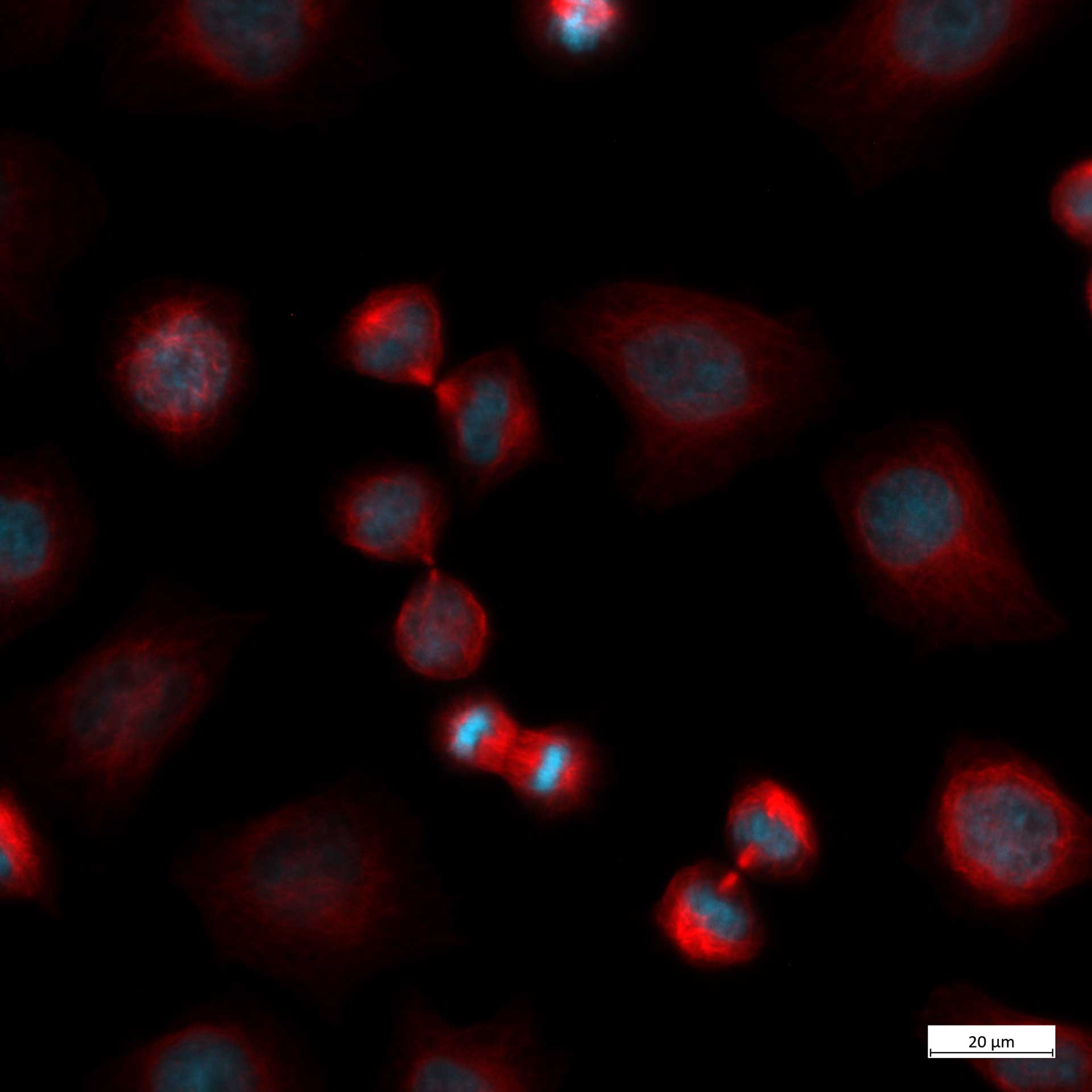
HeLa Kyoto cells, multichannel fluorescence image

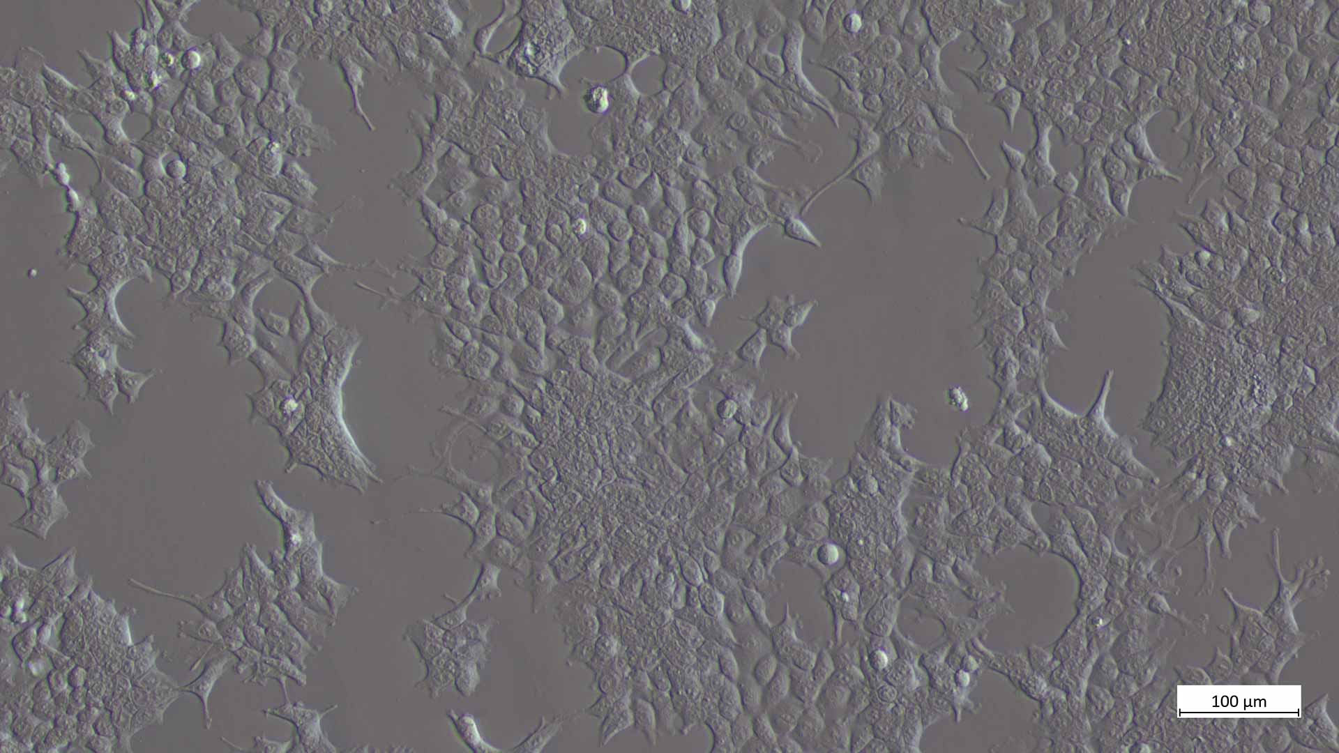
Cos7 cells, improved Hofman modulation contrast


Welding joint. Brightfield, EC Epiplan 5×/0.13


Hardness crack C60 Steel, brightfield, EC Epiplan 20×/0.4


Lithium ion battery. Brightfield, EC Epiplan 20×/0.4


Copper, brightfield, EC Epiplan-Neofluar 10×/0.25


Cast Iron Analysis – image segmentation step


Layer Thickness Measurement – automatic detection of a layer


실체·줌 현미경 어플리케이션 미리보기
-


Fluorescence: Whole mouse embryo


Brightfield for transparent, high-contrast and dyed specimens and to display contours and profiles: Daisy, PlanApo S 1.5x objective, Magnification: 150×


Two-color fluorescence image of drosophila embryo


Human cells identified to be isolated and transported to the PCR tube

Image courtesy of Aalen University Image courtesy of Aalen University Inspection control of coalbrush with Axio Zoom.V16.

Detail of Lithium Ion Battery

Wafer 112x
-


Mouth parts of the common housefly - Transmitted light with VisiLED HCT contrast stage. Lateral darkfield


Mouth parts of the common housefly - Transmitted light with VisiLED HCT contrast stage. Brightfield


SMD-Board with white solder resist. Reflected light with different light guides. Linear slit light for lateral grazing light.


Embryonic stage of a squid, darkfield


Diatoms, transmitted light brightfield


Ofan embryo, brightfield, oblique illumination ("negative“ relief contrast)


PCB component, reflected light darkfield, magnification: 7.5x


PCB component, reflected light darkfield, magnification: 150x (total magnification through the eyepieces)


Daisy (Bellis perennis), transmitted light brightfield, magnification: 150x (visual magnification with 10x eyepieces)
-


Butterfly wing in brightfield


Radish seeds in darkfield


Root tips in brightfield


Spinal cord of feline in brightfield


Royal fern - sori and sporangia acquired with reflected light, darkfield


Poppy seed acquired with reflected light, brightfield


Chip acquired with oblique reflected light

Microfluidics acquired with reflected light


디지털 현미경 어플리케이션 미리보기
-

Electronics: Inspection and failure check of PCBs (Printed Circuit Boards) and SMDs (Surface-Mounted Devices)


Research and Development: Analyzes samples in materials science and studies surfaces at the micro or nano level


Medical Devices: Inspection and failure check of implants, syringes, orthopedic components, and other parts


Automotive Industry: Inspects automotive parts, such as seals and valves, for defects and ensures precision in screws and joints
-


Laser weld, coaxial brightfield illumination, objective: 1.6x, magnification: 32x


Laser shots in airbag cover, full ringlight illumination, extended depth of field (EDF), magnification: 259x


Exhaust residues on outlet valve, segmented ringlight illumination, extended depth of field (EDF), magnification: 45x


Coil wire soldered to contact pin, segmented ringlight illumination, extended depth of field (EDF), magnification: 80x


