
여러분의 파트너
ZEISS Microscopy
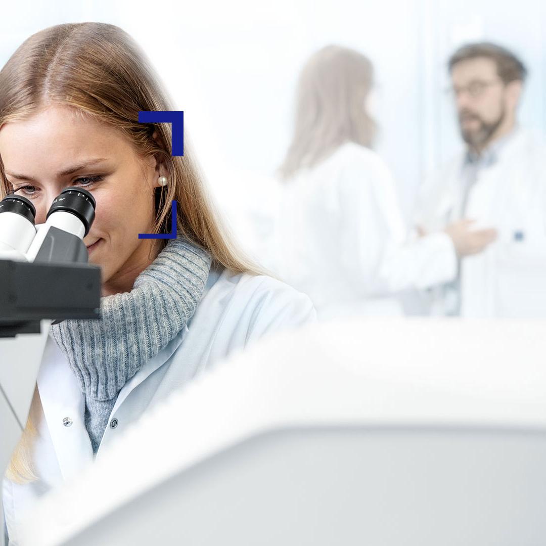
가장 진보한 현미경 솔루션
ZEISS 는 업계를 선도하는 현미경 회사로서, 생명 과학과 재료 연구, 교육 및 임상 실험에 최상의 솔루션과 서비스를 제공합니다. 신뢰할 수 있는 ZEISS 솔루션은 전 세계의 원료 탐구 및 가공, 첨단 기술 산업의 제조 및 조립 분야에서 사용됩니다.
여러분의 어플리케이션에 따라 이상적인 솔루션을 제공하는 광학, 공초점, 전자 및 X선 현미경을 선택하세요!